Recovery & Follow-up
Contents
Recovery includes active monitoring for new neurologic symptoms, bleeding, seizure, wound problems, and changes in blood flow. Direct bypass flow is immediate, indirect collateral growth takes months, and new focal symptoms after surgery need urgent assessment.
Follow-up should distinguish low-flow ischemia, thrombosis or embolism, regional hyperperfusion, bypass patency, indirect collateral maturation, untreated-territory progression, and functional or cognitive recovery.
Imaging gallery
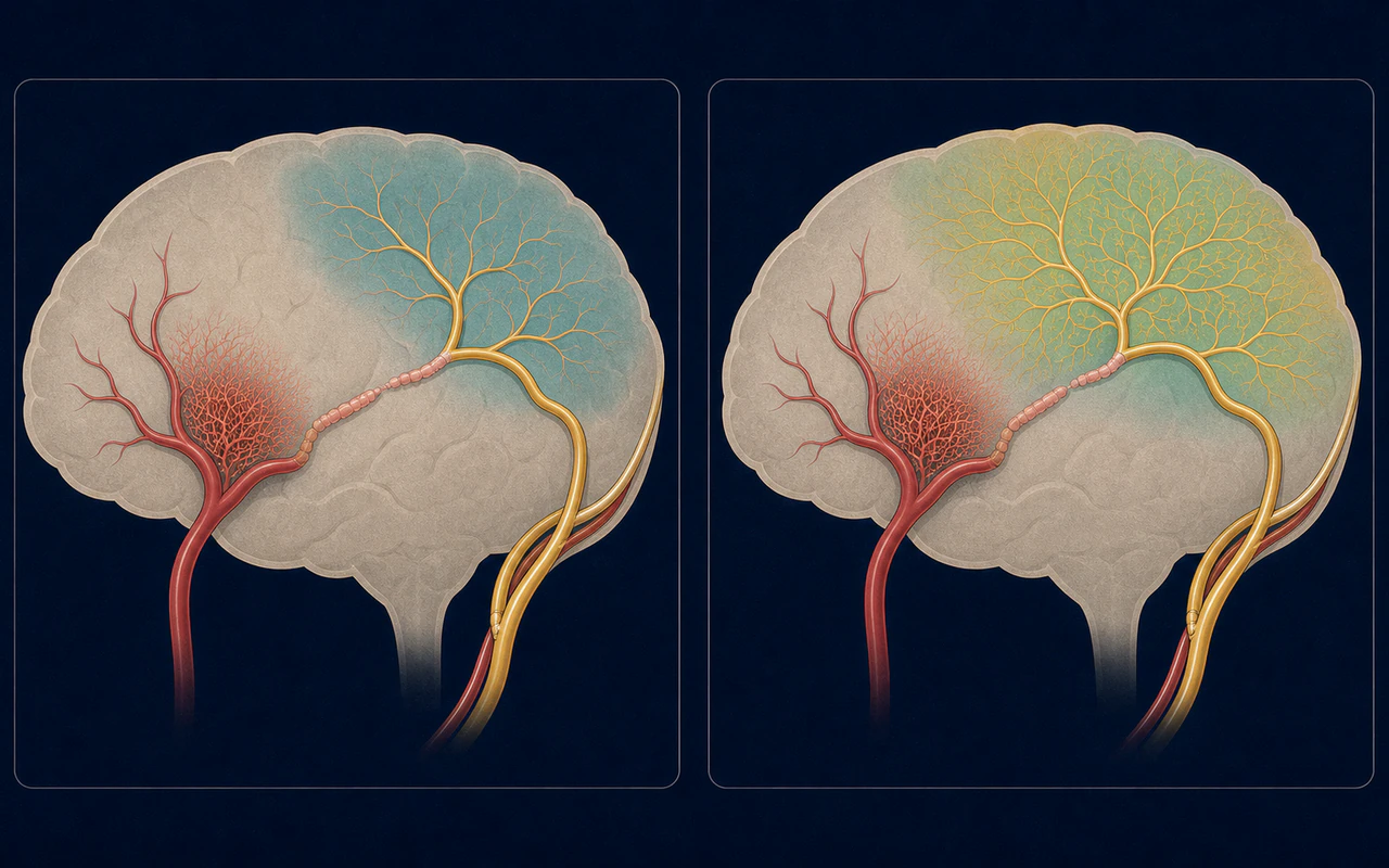
AI-generated educational schematic. Not a real medical image or an exact anatomical representation.
Follow-up may assess direct bypass patency, indirect collateral maturation, perfusion change, and untreated vascular territories.
Hospital recovery
Section titled “Hospital recovery”Protocols differ by center and operation. Common priorities include frequent neurologic checks, pain and nausea control, hydration, oxygenation, temperature, blood pressure, and avoidance of major carbon-dioxide changes. Imaging may be obtained to assess the brain and bypass. The exact targets are patient-specific because both too little and too much regional blood flow can be harmful. [1]
After a direct bypass, flow is present immediately, but the brain must adapt to a new distribution. Cerebral hyperperfusion syndrome is one possible postoperative concern. Ischemic events can also occur from low flow, thrombosis, embolism, or competing circulation. Japanese guidance emphasizes careful postoperative blood-pressure control while considering the risk of ischemia in other territories. [2]
At home
Section titled “At home”Discharge instructions should specify wound care, activity limits, medicines, hydration, headache expectations, emergency symptoms, and who to contact. Fatigue is common after hospitalization and brain surgery, but worsening or focal symptoms should not be assumed to be normal recovery. Return to school, work, driving, exercise, and travel should follow the treating team’s timeline.
Follow-up imaging
Section titled “Follow-up imaging”Direct bypass patency may be assessed with Doppler, CTA, MRA, or angiography depending on the clinical question. Indirect collateral development usually takes months and may be evaluated with MRA or catheter angiography. Perfusion testing can help assess hemodynamic change. Long-term imaging is also used to monitor untreated territories and disease progression. [3]
Rehabilitation and long-term function
Section titled “Rehabilitation and long-term function”Physical, occupational, speech, cognitive, or school-based rehabilitation may be needed after stroke or surgery. Follow-up should address headaches, seizures, cognition, mood, and participation, not only vessel images.
Clinical detail
Section titled “Clinical detail”An excellent angiographic result does not automatically equal complete neurologic recovery, and symptoms do not always map directly to bypass appearance. Outcomes should combine clinical events, function, cognition, perfusion, and vascular imaging.